Standing Apart from the Competition
- "Standing Apart from the Competition" was originally published in the January 2024 health and wellness issue of COMO Magazine.
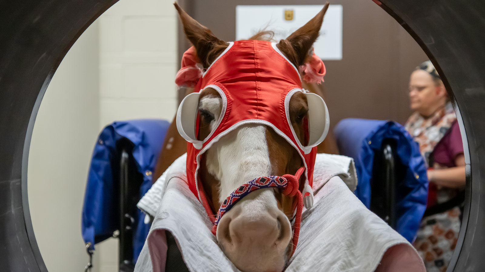
The new standing CT scanner at Clydesdale Hall brings new research and learning opportunities.
In June 2023, The University of Missouri’s College of Veterinary Medicine added a standing CT scanner to Clydesdale Hall. The new machine and improvements to the building were funded through a combination of donor gifts and an endowment program. The standing CT is the first in the region and offers a variety of diagnostic and research possibilities.
“We are always looking for ways to improve our ability to diagnose problems early and find affordable solutions. We also have a lot of innovative clinicians and they tend to be interested in and find other innovative people,” says Joanne Kramer, DVM, MS, DACVS, a teaching professor at the College of Veterinary Medicine. “We were lucky enough to find an innovative company with an engineer who thought outside the circle.”
Traditional CT scanners require a patient to lie down as they are inserted into the machine. When it comes time for animals to undergo such imaging, patients are sedated under general anesthesia. Not only is general anesthesia considered risky for large animals, the procedure requires more staff and more time.
“This new technology allows us to minimize these obstacles and get horses the imaging they need so that we can get a more accurate diagnosis and can recommend more effective treatment,” Kramer explains.
The scanner also allows the animal patient to be standing, which has the added benefit of showing detailed imaging of a joint in its more natural position while also bearing weight on problem areas.
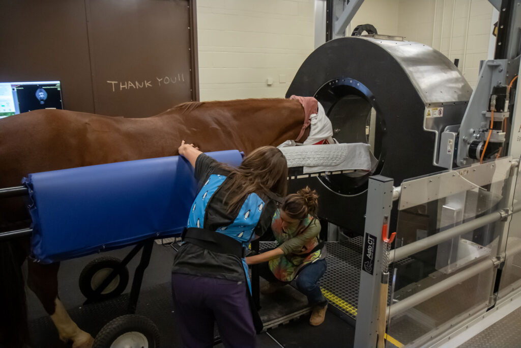
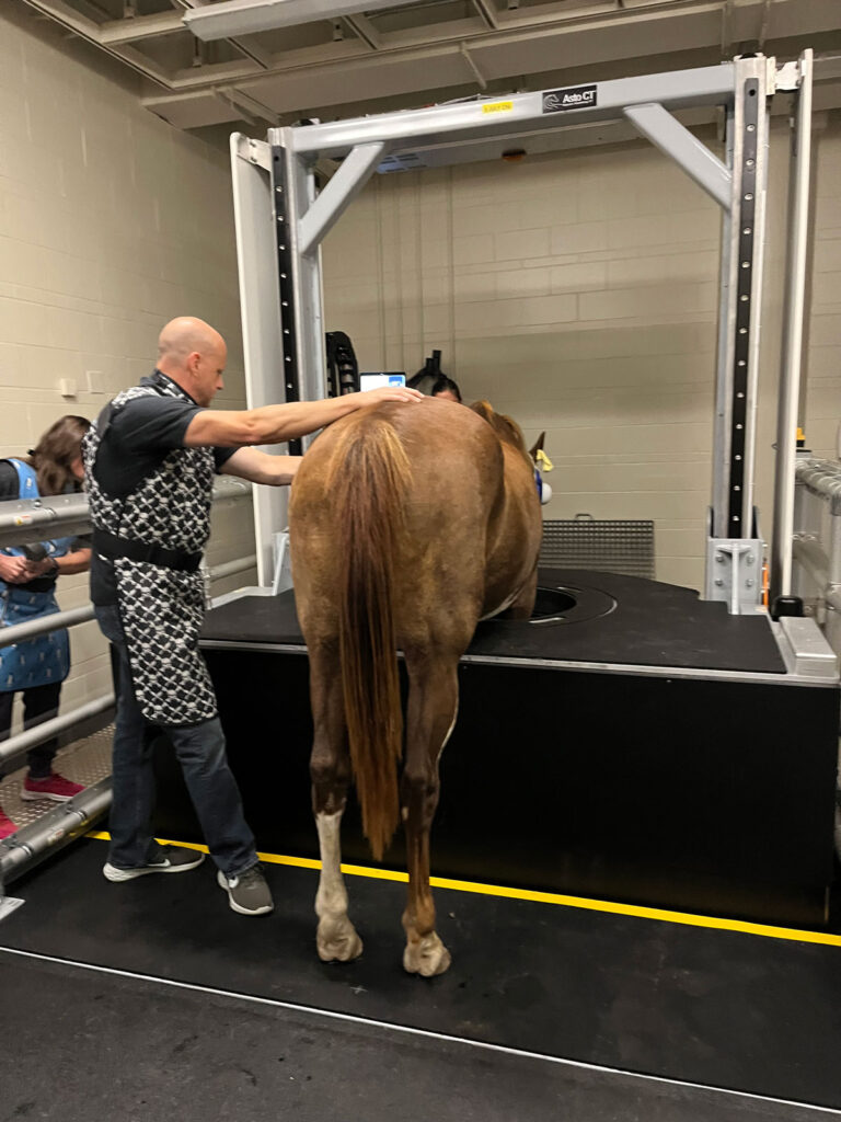
“A standing CT scan can more accurately show the exact orientation of a fracture,” Kramer adds. “Some of the fractures we have seen on standing CT can be treated with bone screws or plates. Knowing the exact orientation enables us to place the screw or plate in the place it can work the best, giving the horse the best chance at making a good recovery.”
The technology can also be used to identify issues before traditional radiography can detect them.
“We have been able to identify more familiar problems like osteoarthritis at an earlier stage than we’ve been able to detect previously,” she says. “This allows us to start treatment at an earlier stage.”
The technology will also be used for research. The lack of intensive prep will allow researchers to do more scans and get more information quickly. She expects that the increased research opportunities will increase enrollment as well.
“We are exploring the possibilities for early detection of fracture risk in racehorses,” she adds. “There are also many important anatomical structures involved in equine lameness and lameness that we can see in more detail and view from more perspectives.”
The technology also allows for more in-depth teaching opportunities. Once mysterious structures can be viewed from a variety of angles, offering students a more elaborate understanding.
“Sometimes the ability to see something from a different perspective helps you lock into the essence of the problem and opens up new ways to think about it,” Kramer explains. “We’re looking at how we can understand and teach things more from that perspective.”
The new technology positions Mizzou’s clinic as a regional leader, servicing not only Missouri but also the region and surrounding states. The clinic’s strategy plan points out that Columbia is centrally located and will provide increased diagnostic access for horses as far away as Kentucky and Wisconsin.
“We hope to create an everyday, everyone CT philosophy,” she said, noting the program’s immediate and future goals. “If a CT scan is indicated we want to be able to offer it to any case that needs it. We believe this technology should and will benefit almost all cases.”


